Wednesday, May 07, 2008

About the site

This material is to assist you with studying for lab. The BEST way to study is to use this as a supplement when you are away from the lab. To do well, you need to also be able to identify these things (slides, display, dissections) on the exam. That means that it is IMPORTANT that you complete all of the dissections, and go into the lab as much as possible so that you can identify the materials when they are not on a computer screen.

GOOD LUCK!!

Tuesday, November 28, 2006

Addition to Annelida 11/28/06


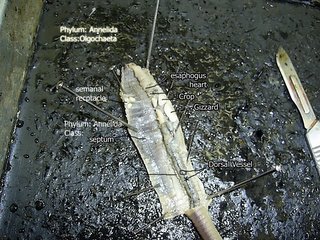
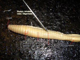
The thing to remember about the earthworm dissection is that it is Class Oligochaeta. Also, try to remember the structures in order as they go down the length of the worm.

2 pairs of nephridia (excretion) can be found per segment.

Friday, November 17, 2006

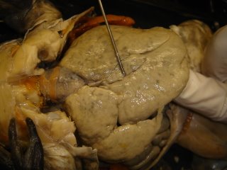
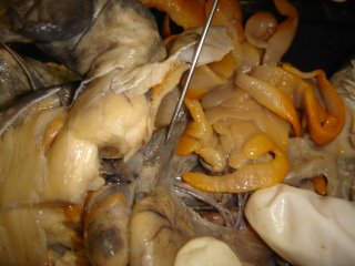
11-16-06 Frog Dissection


Nostrils, Eyes (Shut) and Tympanic membrane on each side (for hearing)


Better view of the nostrils...


Tongue and Eustachian tubes


Eustachian tube


Glottis


Internal nares


Maxillary teeth


Esophagus


Gluteus


Triceps femoris


Semimembranosus


Gastrocnemius


Peroneus


Ileofibularis


Achilles tendon


gracilis major


Triceps femoris


Sartorius


?


Gastrocnemius


Rectus abdominis


Pectoralis


Mylohyoid


Heart


Liver


Stomach


Gall bladder


Small intestine


Large Intestine


Mesentery (membrane)


Spleen


?


Large Intestine


Fat body


leafy stuff beindgheld up is ovary


kidney


Urinary Bladder


Oviduct


Frog is Phylum Chordata, Subphylum Vertebrata, Class Amphibia

11-16-06 Slides


This looks like the fish blood. The way that you can tell the difference between the fish and human blood is that the fish blood has a nucleus, and the human blood does not.


which makes this human blood


sickle cell anemia. Caused by one wrong amino acid on one of the Beta chains of the hemoglobin. Easy to identify because some of the blood cells are sickle shaped.


teased smooth muscle.
One nucleus. Long, thin, tapered.


striated


looks like cardiac muscle


motor neurons


cartilage. You can see the chondrocytes in the matrix (made up of collagen and proteins)


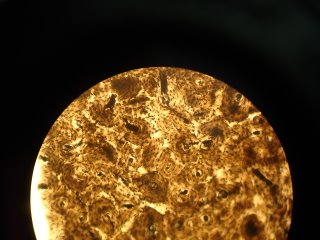
Bone. The spiral things are Haversian systems (Osteon). The center is the Haversian canal. Layers= lamellae. Layers are connected by canalicuti


columnar epithelial


cuboidal epithelial


stratified epithelial (skin)


cuboidal


columnar


columnar


stratified